Saturday, 16 April, 2:00pm-5:45pm
Organizer
- El-Sayed H. Ibrahim (University of Michigan, Ann Arbor, USA)
Co-presenters
- Leon Axel (New York University, New York, USA)
- Choukri Mekkaoui (Harvard University, Boston, USA)
- Dimitri Metaxas (Rutgers University, New Jersey, USA)
- Ralph Sinkus (King’s College London, London, UK)
- Ismail Ben Ayed (School of Superior Technology (ETS), Montreal, Canada)
- Ahmed S. Fahmy (Cairo University, Egypt; Diagnosoft Inc, USA)
Topic and background
The cardiovascular disease is the leading cause of death worldwide with huge associated cost (Billions of dollars) for prevention and treatment. Magnetic resonance imaging (MRI), with its unprecedented capabilities, provides valuable information about the heart condition, including measures of global and regional heart function. Although global measures of cardiac function, e.g. ejection fraction, allow for general evaluation of the heart condition, regional cardiac function measures, e.g. myocardial strain, torsion, and stress, provide information about early heart modeling before the development of cardiac dysfunction and associated changes in the global measures. Therefore, these measures allow for early intervention, efficient treatment planning, and accurate monitoring of disease progression or recession.
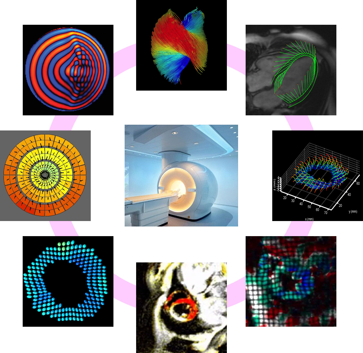
Different MRI techniques have been developed over the past 25 years for measuring heart mechanics and assessing regional cardiac function. These techniques include conventional cine and myocardial tagging (spatial modulation of magnetization (SPAMM) and complementary SPAMM (CSPAMM)) techniques, harmonic-phase (HARP) analysis, strain-encoding (SENC), displacement encoding with stimulated echoes (DENSE), tissue phase mapping (TPM), cine cardiac feature tracking, and magnetic resonance elastography (MRE). Further, the recent advancements in in vivo MRI diffusion tensor imaging (DTI) of the heart allowed for better understanding of the cardiac myofiber structure and its effect on the heart function. These advanced MRI techniques provide different types of MRI images that need special postprocessing and analysis algorithms to efficiently extract accurate and meaningful information about the heart function. Noticeably, each of these techniques has its own advantages and limitations, and different techniques have been used in different clinical and research studies.
Description
This tutorial is presented by world experts in the field from top institutions worldwide, including Dr. El-Sayed Ibrahim, the author of a 2-volume book on the tutorial subject. The tutorial is divided into different presentations, each focusing on one category of the mechanical cardiac imaging techniques listed in the previous paragraph. The first presentation provides an overview of the whole tutorial, different techniques to be covered, historical background about the development of these techniques, and how they are related to each other. Each of the following presentations starts by introducing the basic principles of the imaging techniques covered in the presentation, followed by description of the different development stages of each technique, practical implementation, application examples, and advantages and limitations. The last presentation discusses clinical applications of the covered techniques, techniques’ similarities and differences, current technical challenges, and future development trends. After the first and second parts of the tutorial, there will be a 15-minute ‘meet with the speakers’ event for one-on-one discussions. The tutorial outline, brief description of each presentations, and list of relevant references will be distributed to the audience.
Outline
- Heart Mechanics by Magnetic Resonance Imaging: Past, Present, and Future
- Heart physiology and cardiovascular disease
- The structure-function relationship
- Mechanical heart modeling
- Overview of different MRI techniques for evaluating the heart function
- The Cardiac Structure-Function Relationship: An MRI Perspective
- Myocardial fiber structure and orientation
- Fundamentals of diffusion tensor imaging (DTI)
- Cardiac DTI in Practice
- Myofiber structure and heart function
- Cine Cardiac Magnetic Resonance Image Analysis
- Heart segmentation (Level sets, Graph cuts, Tracking methods)
- Cardiac motion recognition (Support vector machines, Intensity distribution techniques)
- Feature tracking analysis
- Tagged Cardiac Magnetic Resonance Image Analysis
- Basics of Myocardial Tagging
- Optical flow techniques
- Sinusoidal analysis
- Sparsity and machine learning
- Deformable models
- Myocardial Strain Analysis: HARP and SENC
- Harmonic phase (HARP) analysis: the basics
- Real-time HARP
- 3D HARP analysis
- Strain encoding (SENC): the basics
- Real-time SENC
- Composite SENC
- Displacement- and Velocity-Encoding Cardiac Magnetic Resonance Image Analysis
- Displacement encoding with stimulated echoes (DENSE): the basics
- Stimulated echo (STEAM) imaging
- Motion recovery and strain estimation
- Tissue phase mapping (TPM): the basics
- Signal phase encoding
- Myocardial velocity measurement
- Cardiac Magnetic Resonance Elastography (MRE)
- Basic principles of MRE
- Motion equation and mechanical wave propagation
- Reconstruction of elasticity maps
- Cardiac MRE
- Technical Challenges, Clinical Applications, and Future Trends
- Similarities and differences among different techniques
- Technical challenges
- Clinical applications
- Future development trends
Audience
This tutorial is of importance to anyone working on cardiac imaging. The focus on this tutorial is on MRI due to the unprecedented level of details about the heart function that MRI provides compared to other imaging modalities. Nevertheless, some of the covered analysis techniques can be generalized to other imaging modalities. This tutorial is the first to group such important and advanced cardiac functional analysis topics in one session, summarizing the efforts that have been made over the past 25 years to develop these techniques and implement them on clinical applications. With the contents and organization of the topics covered in the different presentations in the tutorial, this tutorial is suitable for audience with either engineering or medical background, as well as with different experience levels from beginners to experts. Hardcopy materials, including list of references relevant to the covered topics, will be provided to the audience for the potential of more thorough readings on these topics. By the end of this tutorial, the audience will obtain comprehensive and up-to-date knowledge about state-of-the-art MRI techniques for evaluating the heart function and mechanical properties, and will be able to identify the advantages, limitations, and suitable clinical applications of each technique.