Clinical Day
Medical imaging plays a major role in the clinical decision-making process in oncology. Until recently, that role has been limited to radiological diagnosis and staging. However, quantitative markers have been shown to be relevant for grading and genotyping of the tumor. These markers can be derived from routine clinical images non-invasively, as a set of intensity and texture based features generally termed Radiomics. Radiomic features have illustrated an impact not only in pathological and genotypic diagnosis of the tumor, but also in radiation treatment planning, understanding effects of chemotherapy etc., paving its way to become a critical entity in oncology. The main purpose of the workshop is to provide a stimulating environment for an in-depth discussion to understand the clinical and research problems to find future relevant solutions in radiomics workflow and its clinical translation.
-

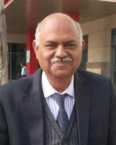
Dr. Rakesh K Gupta, MD
Fortis Memorial Research
India
Biography
Dr. Rakesh Gupta is the Head and Director of Radiology and Imaging in Fortis Memorial Research Institute, Gurgaon, India. He is a neuroradiologist with more than 36 years of experience in clinical neuroimaging and research related to neuroscience studies. He has been involved in teaching and research, and has helped develop various software for clinical applications in neuroscience. He is on the editorial board of AJNR, JMRI and Neuroradiology and has been awarded fellowships of ISMRM, National Science Academy, and Academy of Medical Science (India). He has published more than 475 papers in the peer-reviewed international and national journals, and has mentored 15 PhDs and several MD and DM students.Abstract
Brain tumours are highly heterogeneous and carry poor prognosis and rank among the top 10 causes of cancer deaths. MRI is a routinely performed for patients who are suspected of brain tumours and is the main stay in its diagnosis and treatment response assessment. A number of advanced MRI techniques like diffusion-based imaging, perfusion imaging and MR spectroscopy are now routinely used in the evaluation of brain tumours. The application of radiomics has been initiated in clinical oncology due to its ability to analyse the combination of numerous quantitative features provides the possibility to unravel the underlying pathophysiology that is hard to be perceived by radiologists’ eyes and avoid subjective misreading. The general workflow of radiomics involves several discrete steps: imaging, segmentation, feature extraction, feature selection, machine learning, and validation. In this presentation, we intend to demonstrate the possible role of radiomics in brain tumor and how it may support in clinical decision making in overall management of the disease in future. The current challenges associated with radiomics will also be discussed. -

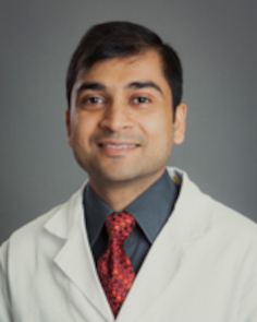
Dr. Suyash Mohan
Perelman School of Medicine at the University of Pennsylvania
USA
Biography
Dr. Mohan is a physician-scientist with a research focus on clinical and translational applications of advanced neuroimaging techniques in the development of noninvasive imaging-based biomarkers for assessing brain tumor metabolism and response to therapy. His current NIH/NCI funded clinical trial is utilizing synergy of advanced metabolic imaging, computational modelling, with interdisciplinary collaborations and industrial partnership to transform the treatment of patients with brain tumors. Dr. Mohan is the recipient of numerous awards and honors and has been the brand ambassador for societies like the International Society for Magnetic Resonance in Medicine (ISMRM) and the American Society of Neuroradiology (ASNR). He was awarded the 2018 Anne G. Osborn ASNR Outreach Professorship to South Africa and will be the RSNA International Visiting Professor to Kazakhstan in 2022. Dr. Mohan is a passionate educator and was the recipient of the Wallace T. Miller Teaching Award for Teaching Excellence in 2013, and then the ‘2015 Teaching Award’, for ‘Excellence in Teaching’ at the University of Pennsylvania. Most recently, he was awarded the Association of University Radiologists (AUR), 2019 Outstanding Teacher Award. Dr. Mohan has made important scientific contributions and has published over 125 peer-reviewed articles, numerous book chapters, presented over 200 abstracts in national and international meetings, and has delivered over 300 lectures locally, nationally, and internationally. Dr. Mohan has had a number of roles in national organizations, particularly the American Society of Neuroradiology (ASNR) and the Radiological Society of North America (RSNA), and was recently appointed as the Assistant Editor for Radiographics. He has focused interest in advancing neuro-oncologic imaging and research, teaching and education, academic mentoring of trainees and junior faculty, as well as in multidisciplinary collaborations.
Abstract
Glioblastoma is the most common and most aggressive form of primary brain tumor in adults. Despite aggressive multimodal treatment, its prognosis remains poor. Conventional imaging is limited in delineating the full extent of this extremely infiltrative tumor, and ‘what we cannot see is what we cannot treat’. In this talk we will discuss some of the newer advances that we have seen in the field of neuro-oncologic imaging, specifically for precision diagnostics, review novel imaging based biomarkers and see how these techniques are playing an increasingly meaningful role in informing clinical care and clinical trials.
Machine learning (ML) integrated with these neuro-oncologic imaging techniques has introduced new perspectives in precision diagnostics, through radiomics and radiogenomics. In this talk, we will also review practical computational perspectives of some of these radiomic based noninvasive-in-vivo biomarkers for allowing personalized treatments for these brain tumor patients and discuss future directions. -

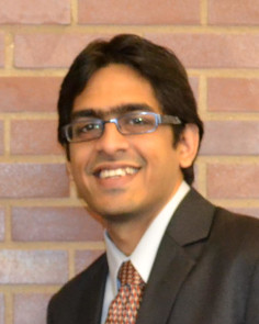
Dr. Satish Viswanath
Case Western Reserve UniversityUSA
Abstract
Developing artificial intelligence (AI) schemes to assist the clinician towards enabling precision medicine requires “unlocking” embedded information captured by different data modalities, in an intuitive and generalizable fashion. The research in my group focuses on developing novel computational imaging features (termed “radiomic” features) together with histology or molecular data for disease characterization and treatment response evaluation in vivo. Towards this, we have designed unique tools that can capture biologically relevant and clinically intuitive measurements from routinely acquired imaging (MRI, CT, PET) or digitized images of tissue specimens. In addition to developing approaches to ensure these models generalize tio new unseen data, we have also evaluated their repeatability across imaging parameters and reproducibility across institution- or scanner-specific variations. Problems being addressed by us include: (a) predicting response to treatment to identify optimal therapeutic pathways, as well as (b) evaluating therapeutic response to guide follow-up procedures; in the context of colorectal cancers and digestive diseases.

