Wednesday, 13 April, 8:30am-12:15pm
Organizers
- The International Skin Imaging Collaboration
- Allan Halpern (Clinical Leader) (Memorial Sloan Kettering Cancer Center, New York, USA)
- Brian Helba (Technical Leader) (Kitware, New York, USA)
- David Gutman (Technical Leader) (Emory University, Atlanta, USA)
- Noel Codella (Image Analysis) (IBM, New York, USA)
- Emre Celebi (Image Analysis) (Louisiana State University, Shreveport, Louisiana)
Challenge website
Challenge abstract
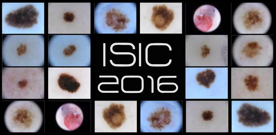
Skin cancer is a major public health problem, with over 5 million newly diagnosed cases in the United States each year. Melanoma is the deadliest form of skin cancer, responsible for over 9,000 deaths each year. As pigmented lesions occurring on the surface of the skin, melanoma is amenable to early detection by expert visual inspection. It is also amenable to automated detection with image analysis. Given the widespread availability of high-resolution cameras, algorithms that can improve our ability to screen and detect troublesome lesions can be of tremendous value. As a result, tremendous interest has grown, as many centers have begun their own research efforts on automated analysis. However, a centralized, coordinated, and comparative effort across institutions has yet to be implemented.
The International Skin Imaging Collaboration (ISIC) is an international effort to improve melanoma diagnosis. This challenge leverages a database of skin images from the ISIC data archive, which contains over 10,000 dermoscopic images collected from leading clinical centers internationally, acquired from a variety of devices used at each center. The images are screened for both privacy and quality assurance. The associated clinical metadata has been vetted by recognized melanoma experts. Broad and international participation in image contribution is designed to insure a representative clinically relevant sample.
The overarching goal of the challenge is to develop image analysis tools to enable the automated diagnosis of melanoma from dermoscopic images. The challenge will be divided into sub-challenges on lesion segmentation, lesion dermoscopic feature detection and lesion classification.
Other challenges
 |
Cancer Metastasis Detection in Lymph Nodes (CAMELYON) |
 |
Analysis of Images to Detect Abnormalities in Endoscopy (AIDA-E) |
 |
Lung Nodule Analysis (LUNA) |
