Keynotes speakers

Stéphane Mallat
Professor, Collège de France, PSL University, France
Wednesday 14, April
Biography
Stéphane Mallat received the Ph.D. degree in electrical engineering from the University of Pennsylvania, in 1988. He was then Professor at the Courant Institute of Mathematical Sciences, until 1994. In 1995, he became Professor in Applied Mathematics at Ecole Polytechnique, Paris and Department Chair in 2001. From 2001 to 2007 he was co-founder and CEO of a semiconductor start-up company. From 2012 to 2017 he was Professor in the Computer Science Department of Ecole Normale Supérieure, in Paris. Since 2017, he holds the “Data Sciences” chair at the Collège de France.
Stéphane Mallat’s research interests include machine learning, signal processing, and harmonic analysis. He is a member of the French Academy of sciences, of the Academy of Technologies, a foreign member of the US National Academy of Engineering, an IEEE Fellow and an EUSIPCO Fellow. In 1997, he received the Outstanding Achievement Award from the SPIE Society and was a plenary lecturer at the International Congress of Mathematicians in 1998. He also received the 2004 European IST Grand prize, the 2004 INIST-CNRS prize for most cited French researcher in engineering and computer science, the 2007 EADS grand prize of the French Academy of Sciences, the 2013 Innovation medal of the CNRS, the 2015 IEEE Signal Processing best sustaining paper award, and the 2018 IEEE Freidrich Gauss educational award.
Abstract: A Mathematical View of Deep Neural Networks
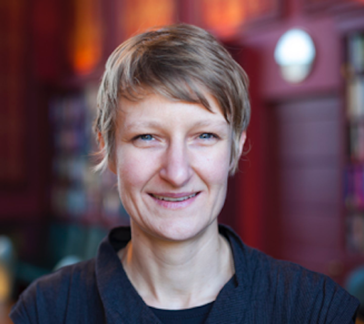
Carola-Bibiane Schönlieb
Professor, University of Cambridge, United Kingdom
Thursday 15, April
Biography
Carola-Bibiane Schönlieb is Professor of Applied Mathematics at the Department of Applied Mathematics and Theoretical Physics (DAMTP), University of Cambridge. There, she is head of the Cambridge Image Analysis group, Director of the Cantab Capital Institute for Mathematics of Information, and Director of the EPSRC Centre for Mathematical and Statistical Analysis of Multimodal Clinical Imaging. Since 2011 she is a fellow of Jesus College Cambridge and since 2016 a fellow of the Alan Turing Institute, London.
Her research has been acknowledged by scientific prizes, among them the LMS Whitehead Prize 2016, the Philip Leverhulme Prize in 2017, and the Calderon Prize 2019, and by invitations to give plenary lectures at several renowned applied mathematics conferences, among them the SIAM conference on Imaging Science in 2014, the SIAM conference on Partial Differential Equations in 2015, the SIAM annual meeting in 2017, the Applied Inverse Problems Conference in 2019 and FOCM 2020.
Carola graduated from the Institute for Mathematics, University of Salzburg (Austria) in 2004. From 2004 to 2005 she held a teaching position in Salzburg. She received her PhD degree from the University of Cambridge in 2009. After one year of postdoctoral activity at the University of Göttingen (Germany), she became a Lecturer in at DAMTP in 2010, promoted to Reader in 2015 and promoted to Professor in 2018. Her current research interests focus on variational methods, partial differential equations and machine learning for image analysis, image processing and inverse imaging problems.
She has active interdisciplinary collaborations with clinicians, biologists and physicists on biomedical imaging topics, chemical engineers and plant scientists on image sensing, as well as collaborations with artists and art conservators on digital art restoration.
Abstract: Combining knowledge and data driven approaches to biomedical imaging - getting the best from both worlds
Biography
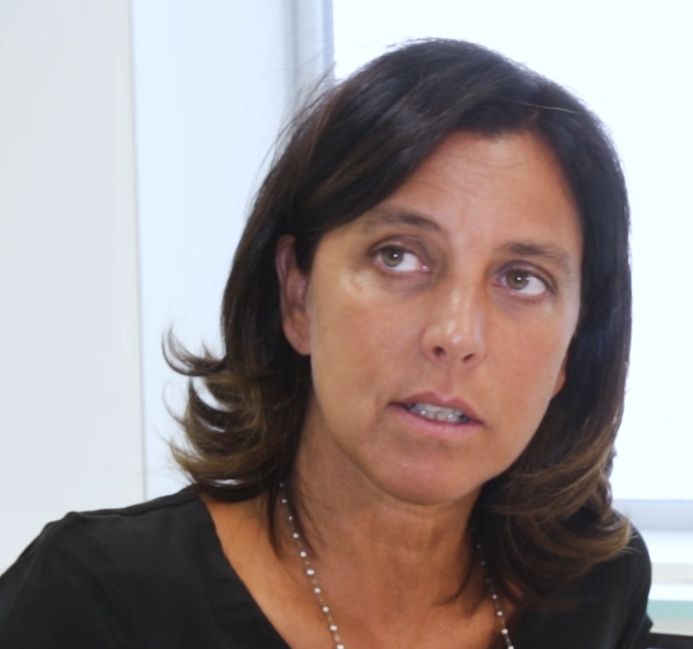
Dr. Valentina Emiliani is a CNRS Research Director at the Vision Institute in Paris where she leads the Photonics Department and the ‘Wave front engineering microscopy’ group. After her PhD in Physics (University La Sapienza, Rome) she worked as a post doc at the Max Born Institute (Berlin) to investigate carrier transport in quantum wire by low temperature scanning near field optical microscopy. Later on, she worked at the European Laboratory for Nonlinear Spectroscopy to lead the research group: ‘High resolution microscopy’, focused on the investigation of light propagation in disordered structure by near field microscopy. In 2002 she moved to Paris at the Institute Jacques Monod and in 2005 she was awarded with the European Young Investigator grant to form the “Wave front engineering microscopy” group at Paris Descartes University. In 2019 she moved the group at the vision Institute in Paris. In 2015 she has been awarded with the Prix “Coups d’élan pour la recherche française” from the Bettencourt-Shueller foundation, in 2017 she has been awarded with the Axa chair “Investigation of visual circuits by optical wave front shaping “ and in 2020 with the ERC advanced grant.
Dr. Valentina Emiliani has pioneered the use of wave-front engineering for neuroscience. In particular, with the ‘Wave front engineering microscopy” group she demonstrated a number of new optical methods for efficient photoactivation of caged compounds and optogenetics molecules, techniques based on computer generated holography, generalized phase contrast and temporal focusing. Her current interest is to use wave front shaping and optogenetics for the investigation of the mechanisms regulating functional connectivity and signal processing across the main visual pathways.
In 2021, she received the « Silver medal » of CNRS.

